New interesting publication by Macrea et. al mentioning the use of NANOSENSORS uniqprobe qp-BioAC:
“Hemostasis requires conversion of fibrinogen to fibrin fibers that generate a characteristic network, interact with blood cells, and initiate tissue repair. The fibrin network is porous and highly permeable, but the spatial arrangement of the external clot face is unknown. Here we show that fibrin transitioned to the blood-air interface through Langmuir film formation, producing a protective film confining clots in human and mouse models. We demonstrated that only fibrin is required for formation of the film, and that it occurred in vitro and in vivo. The fibrin film connected to the underlying clot network through tethering fibers. It was digested by plasmin, and formation of the film was prevented with surfactants. Functionally, the film retained blood cells and protected against penetration by bacterial pathogens in a murine model of dermal infection. Our data show a remarkable aspect of blood clotting in which fibrin forms a protective film covering the external surface of the clot, defending the organism against microbial invasion.”*
The AFM imaging and force measurements mentioned in this article were performed using CB3 of the NANOSENSORS™ uniqprobe qp-BioAC.
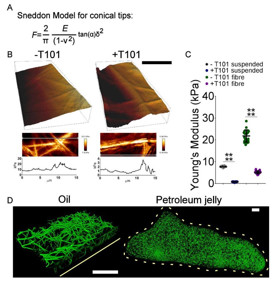
Supplemental Figure. 6. From Macrea et. al “A fibrin biofilm covers blood clots and protects from microbial invasion” Mechanisms and roles of fibrin film. A, Sneddon model used to calculate Young’s Modulus, where F is the force from the force curve, E is Young’s modulus, ν is Poisson’s ratio (0.5), α is the half angle for the indenter (15 degrees for our tips), and δ is the indentation. Note that this equation is only accurate with a half angle of 15 degrees for the first 200nm of indentation. B, Strength of the fibrin film in clots produced with plasma and thrombin with or without T101 (FXIII inhibitor ) investigated using atomic force microscopy (AFM). Fibrin fibres were visible under the film surface and these areas presented with stiffer Young’s modulus than fibrin film suspended between fibres. Grey lines in the zoomed-in images represent Young’s modulus scan area represented in the line force graphs. Scale bar – 2μm. C, Young’s Modulus was calculated for the suspended film and the film supported by fibers with and without T101 by fitting a Sneddon model to all AFM force curves found over the entire area that was imaged. 20 measurements were taken for each condition. **** P<0.0001. D, Clots produced from plasma with thrombin, under a layer of oil or enclosed in a ball of petroleum jelly, to eliminate the air – liquid interface, imaged by LSCM. Solid and dotted yellow lines indicate location of air liquid interface, n=3 experiments. Scale bars – 50μm.
*Fraser L. Macrae, Cédric Duval, Praveen Papareddy, Stephen R. Baker, Nadira Yuldasheva, Katherine J. Kearney, Helen R. McPherson, Nathan Asquith, Joke Konings, Alessandro Casini, Jay L. Degen, Simon D. Connell, Helen Philippou, Alisa S. Wolberg, Heiko Herwald, Robert A.S. Ariëns
A fibrin biofilm covers blood clots and protects from microbial invasion
Journal of Clinical Investigation. 2018;128(8):3356-3368
DOI: https://doi.org/10.1172/JCI98734
Please follow this external link for the full article: https://www.jci.org/articles/view/98734#sd
The article “A fibrin biofilm covers blood clots and protects from microbial invasion” by Fraser L. Macrae et. al is licensed under the Creative Commons Attribution 4.0 International License. To view a copy of this license, visit http://creativecommons.org/ licenses/by/4.0/.