MeCP2 and MBD2 are members of a family of proteins that possess a domain that selectively binds 5-methylcytosine in a CpG context. Members of the family interact with other proteins to modulate DNA packing. Stretching of DNA–protein complexes in nanofluidic channels with a cross-section of a few persistence lengths allows us to probe the degree of compaction by proteins.*
In the article “DNA looping by two 5-methylcytosine-binding proteins quantified using nanofluidic devices” Ming Liu, Saeid Movahed, Saroj Dangi, Hai Pan, Parminder Kaur, Stephanie M. Bilinovich, Edgar M. Faison, Gage O. Leighton, Hong Wang, David C. Williams Jr. and Robert Riehn demonstrate DNA compaction by MeCP2 while MBD2 does not affect DNA configuration. By using atomic force microscopy (AFM), they determined that the mechanism for compaction by MeCP2 is the formation of bridges between distant DNA stretches and the formation of loops.*
Despite sharing a similar specific DNA-binding domain, the impact of full-length 5-methylcytosine-binding proteins can vary drastically between strong compaction of DNA and no discernible large-scale impact of protein binding. The authors of the article demonstrate that ATTO 565-labeled MBD2 is a good candidate as a staining agent for epigenetic mapping.*
For atomic force microscopy (AFM), the authors used a 7,163-bp linear DNA substrate which contains a 1,697-bp methylated CpG-rich region that is flanked by 2,742-bp and 2,724-bp CpG-free regions. For MeCP2, the DNA substrate and the protein were diluted in AFM imaging buffer (HEPES 20 mM, Mg(OAc)210mM, NaCl 100mM, pH 7.5), mixed together and deposited on freshly peeled mica. For MBD2FLsc, the authors describe how they first mixed the protein and DNA and then diluted the sample in AFM buffer before deposition. The final MeCP2 concentration deposited on mica was 7.5nM, and the MBD2FLsc concentration was 14nM. The mica samples were then washed with filtered deionized water and dried with nitrogen.*
NANOSENSORS™ PointProbe® Plus PPP-FMR AFM probes ( ≈2.8N/m) were used to image the sample at a scan resolution of 5.9nm and a scan rate of 3μm/s.*
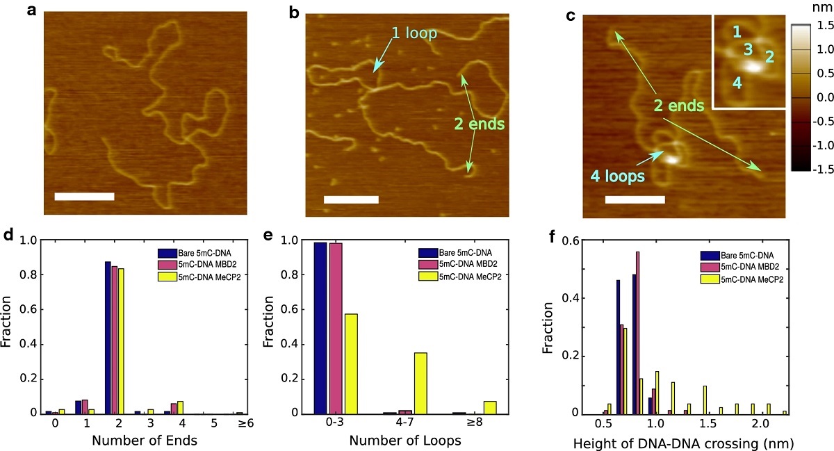
Atomic force microscopy (AFM) of methylated substrates under various conditions. AFM of bare methylated dsDNA oligomer a, the same oligomer with MBD2Flsc b, and with MeCP2 c. Scale bars are 200nm. The green arrows point at ends, and cyan arrows point at loops. The inset in d illustrates the counting method for loops. The distribution of number of free ends (d) and the distribution of number of loops (e) for DNA or DNA–protein complexes was determined from such images (bare DNA N=118, MBD2FLsc N=98, MeCP2 N=108). f Height of isolated DNA–DNA crossings (bare DNA N=52, MBD2FLsc N=68, MeCP2 N=83)
*Ming Liu,
Saeid Movahed, Saroj Dangi, Hai Pan, Parminder Kaur, Stephanie M. Bilinovich,
Edgar M. Faison, Gage O. Leighton, Hong Wang, David C. Williams Jr. and Robert
Riehn
DNA looping by two 5-methylcytosine-binding proteins quantified using
nanofluidic devices
Epigenetics & Chromatin volume 13, Article number: 18 (2020)
DOI: https://doi.org/10.1186/s13072-020-00339-7
Please follow this external link to read the full article: https://rdcu.be/b3iTm
Open Access: The article “DNA looping by two 5-methylcytosine-binding proteins quantified using nanofluidic devices” by Ming Liu, Saeid Movahed, Saroj Dangi, Hai Pan, Parminder Kaur, Stephanie M. Bilinovich, Edgar M. Faison, Gage O. Leighton, Hong Wang, David C. Williams Jr. and Robert Riehn is licensed under a Creative Commons Attribution 4.0 International License, which permits use, sharing, adaptation, distribution and reproduction in any medium or format, as long as you give appropriate credit to the original author(s) and the source, provide a link to the Creative Commons license, and indicate if changes were made. The images or other third party material in this article are included in the article’s Creative Commons license, unless indicated otherwise in a credit line to the material. If material is not included in the article’s Creative Commons license and your intended use is not permitted by statutory regulation or exceeds the permitted use, you will need to obtain permission directly from the copyright holder. To view a copy of this license, visit http://creativecommons.org/licenses/by/4.0/.