Traditional Joule dissipation omnipresent in today’s electronic devices is well understood while the energy loss of the strongly interacting electron systems remains largely unexplored. Twisted bilayer graphene (tBLG) is a host to interaction-driven correlated insulating phases, when the relative rotation is close to the magic angle (1.08∘). *
In the article “Energy dissipation on magic angle twisted bilayer graphene” Alexina Ollier, Marcin Kisiel, Xiaobo Lu, Urs Gysin, Martino Poggio, Dmitri K. Efetov and Ernst Meyer report on low-temperature (5K) nanomechanical energy dissipation of tBLG measured by pendulum atomic force microscopy (p-AFM). *
Owing to high force sensitivity, pendulum geometry Atomic Force Microscopy (p-AFM), oscillating like a tiny pendulum over the surface, is perfectly suited to measure tiny amounts of energy loss.*
The ultrasensitive cantilever tip acting as an oscillating gate over the quantum device shows dissipation peaks attributed to different fractional fillings of the flat energy bands. Local detection allows to determine the twist angle and spatially resolved dissipation images showed the existence of hundred-nanometer domains of different doping. *
Application of magnetic fields provoked strong oscillations of the dissipation signal at 3/4 band filling, identified in analogy to Aharonov-Bohm oscillations, a wavefunction interference present between domains of different doping and a signature of orbital ferromagnetism.*
During the Pendulum AFM energy dissipation measurements series of insulating states of tBLG were detected under ultra high vacuum (UHV) conditions with highly n-doped silicon NANOSENSORS AdvancedTEC ATEC-CONT AFM probes. *
The AFM cantilever with resistivity ρ = 0.01 − 0.02 Ωcm, spring constant k = 0.18 N/m and frequency f0 = 13 kHz was coupled capacitively to the quantum device, and the AFM tip oscillation amplitude A = 1 nm was parallel to the sample surface (pendulum geometry). The sensor was annealed before experiment at 700∘ under UHV conditions for 12h. The process leads to removal of water and weakly bounded molecules from the cantilever surface and the tip. Moreover, the long-term annealing minimizes the amount of the static charges localized at the AFM tip. *

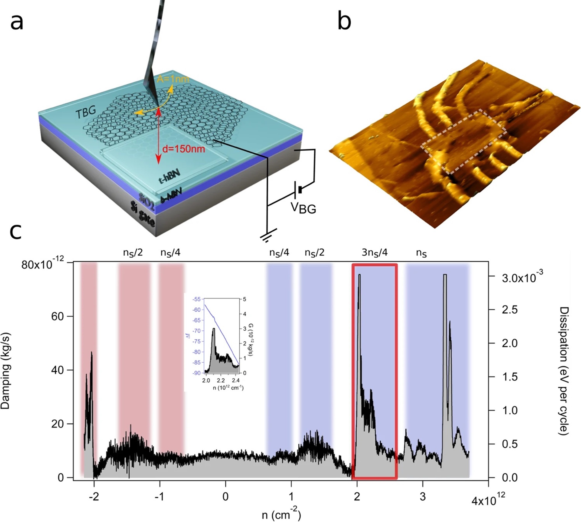
Fig. 1 from Alexina Ollier et al 2023 “Energy dissipation on magic angle twisted bilayer graphene”:
Pendulum atomic force microscope (p-AFM) tip oscillating on top of a twisted bilayer graphene (tBLG) device is measuring energy loss.
a schematics of the measured device. The sample is composed of a p-doped silicon backgate (dark gray), followed by a 300 nm thick silicon dioxide layer (violet) and 10 nm hexagonal boron nitride (hBN, light blue). From the top tBLG is encapsulated with 10nm hBN film. b p-AFM image of the tBLG device, the white dotted rectangle is the device with a size of 4.1 μm × 5.8 μm. In c the typical dissipation (Γ) spectrum versus doping concentration (n) is shown. Different peaks in Γ spectrum are identified as ν = ± 1/4, ν = ± 1/2, ν = ± 3/4, and ν = 4/4 and colored in red and blue for holes and electrons, respectively. The inset shows the corresponding frequency shift Δf(3ns/4) spectrum. The measurement was performed at temperature T = 5 K.
NANOSENSORS AdvancedTEC ATEC-CONT tip-view AFM probes were used.
*Alexina Ollier, Marcin Kisiel, Xiaobo Lu, Urs Gysin, Martino Poggio, Dmitri K. Efetov and Ernst Meyer
Energy dissipation on magic angle twisted bilayer graphene
nature Communications Physics volume 6, Article number: 344 (2023)
DOI: https://doi.org/10.1038/s42005-023-01441-4
The article “Energy dissipation on magic angle twisted bilayer graphene” by Alexina Ollier, Marcin Kisiel, Xiaobo Lu, Urs Gysin, Martino Poggio, Dmitri K. Efetov and Ernst Meyer is licensed under a Creative Commons Attribution 4.0 International License, which permits use, sharing, adaptation, distribution and reproduction in any medium or format, as long as you give appropriate credit to the original author(s) and the source, provide a link to the Creative Commons license, and indicate if changes were made. The images or other third-party material in this article are included in the article’s Creative Commons license, unless indicated otherwise in a credit line to the material. If material is not included in the article’s Creative Commons license and your intended use is not permitted by statutory regulation or exceeds the permitted use, you will need to obtain permission directly from the copyright holder. To view a copy of this license, visit https://creativecommons.org/licenses/by/4.0/.