Ferroic materials are well known to exhibit heterogeneity in the form of domain walls. Understanding the properties of these boundaries is crucial for controlling functionality with external stimuli and for realizing their potential for ultra-low power memory and logic devices as well as novel computing architectures.*
In the article “Infrared nano-spectroscopy of ferroelastic domain walls in hybrid improper ferroelectric Ca3Ti2O7” K. A. Smith, E. A. Nowadnick, S. Fan, O. Khatib, S. J. Lim, B. Gao, N. C. Harms, S. N. Neal, J. K. Kirkland, M. C. Martin, C. J. Won, M. B. Raschke, S.-W. Cheong, C. J. Fennie, G. L. Carr, H. A. Bechtel and J. L. Musfeldt employ synchrotron-based near-field infrared nano-spectroscopy to reveal the vibrational properties of ferroelastic (90∘ ferroelectric) domain walls in the hybrid improper ferroelectric Ca3Ti2O7 . By locally mapping the Ti-O stretching and Ti-O-Ti bending modes, they reveal how structural order parameters rotate across a wall. Thus, they link observed near-field amplitude changes to underlying structural modulations and test ferroelectric switching models against real space measurements of local structure. This initiative opens the door to broadband infrared nano-imaging of heterogeneity in ferroics.*
NANOSENSORS™ Platinum Silicide PtSi-NCH AFM probes were used for the Near-field infrared spectroscopy. Atomic force and piezoforce imaging reveal the different orientations of directional order parameters and domain wall character, providing a physical playground for graph theory. *
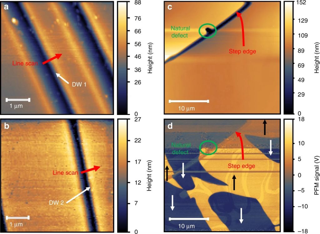
Combining scanning techniques to locate domain walls. a, b Atomic force microscopic (AFM) images of the crystal surfaces showing the two ferroelastic domain walls of interest (at the edges of the dark blue stripes). These ferroelastic walls separate domains of different spontaneous strain and are also 90∘ ferroelectric walls. DW 1 and DW 2 refer to domain walls 1 and 2. Red arrows indicate direction and path of the line scans. The nano-spectroscopic line scans are taken perpendicular to the wall, and the contact angle from one domain to another is 90∘. c AFM topography of a smooth area near an identified surface defect (indicated by a green circle) and step edge of approximately 100 nm height (indicated with a red arrow) compared with d the piezoresponse force microscopic (PFM) image of the same area revealing the placement and orientation of the 180∘ ferroelectric domains, indicated by yellow(+) or blue(−) regions with black or white arrows to indicate the polarization direction. All of these structures are present at room temperature
*K. A. Smith, E. A. Nowadnick, S. Fan, O. Khatib, S. J. Lim, B. Gao, N. C. Harms, S. N. Neal, J. K. Kirkland, M. C. Martin, C. J. Won, M. B. Raschke, S.-W. Cheong, C. J. Fennie, G. L. Carr, H. A. Bechtel and J. L. Musfeldt
Infrared nano-spectroscopy of ferroelastic domain walls in hybrid improper ferroelectric Ca3Ti2O7
Nature Communications volume 10, Article number: 5235 (2019)
DOI: https://doi.org/10.1038/s41467-019-13066-9
Please follow this external link to read the full article: https://rdcu.be/b2rPq
Open Access: The article “Infrared nano-spectroscopy of ferroelastic domain walls in hybrid improper ferroelectric Ca3Ti2O7” by K. A. Smith, E. A. Nowadnick, S. Fan, O. Khatib, S. J. Lim, B. Gao, N. C. Harms, S. N. Neal, J. K. Kirkland, M. C. Martin, C. J. Won, M. B. Raschke, S.-W. Cheong, C. J. Fennie, G. L. Carr, H. A. Bechtel and J. L. Musfeldt is licensed under a Creative Commons Attribution 4.0 International License, which permits use, sharing, adaptation, distribution and reproduction in any medium or format, as long as you give appropriate credit to the original author(s) and the source, provide a link to the Creative Commons license, and indicate if changes were made. The images or other third party material in this article are included in the article’s Creative Commons license, unless indicated otherwise in a credit line to the material. If material is not included in the article’s Creative Commons license and your intended use is not permitted by statutory regulation or exceeds the permitted use, you will need to obtain permission directly from the copyright holder. To view a copy of this license, visit http://creativecommons.org/licenses/by/4.0/.