Amyloidogenesis of α-synuclein (αS) is considered to be a pathological phenomenon related to Parkinson’s disease (PD).
In the publication “Morphological Evaluation of Meta-stable Oligomers of α-Synuclein with Small-Angle Neutron Scattering” Ghibom Bhak, Soonkoo Lee, Tae-Hwan Kim, Ji-Hye Lee, Jee Eun Yang, Keehyoung Joo, Jooyoung Lee, Kookheon Char and Seung R. Paik have studied one particular type of meta-stable αS oligomers (Meta-αS-Os) since they act as a growing unit to exhibit the accelerated amyloid fibril formation in the presence of external stimuli such as shear force10, temperature change11, pH12, and organic solvents13 which are suspected to alter the structure of Meta-αS-Os into a self-associative state.*

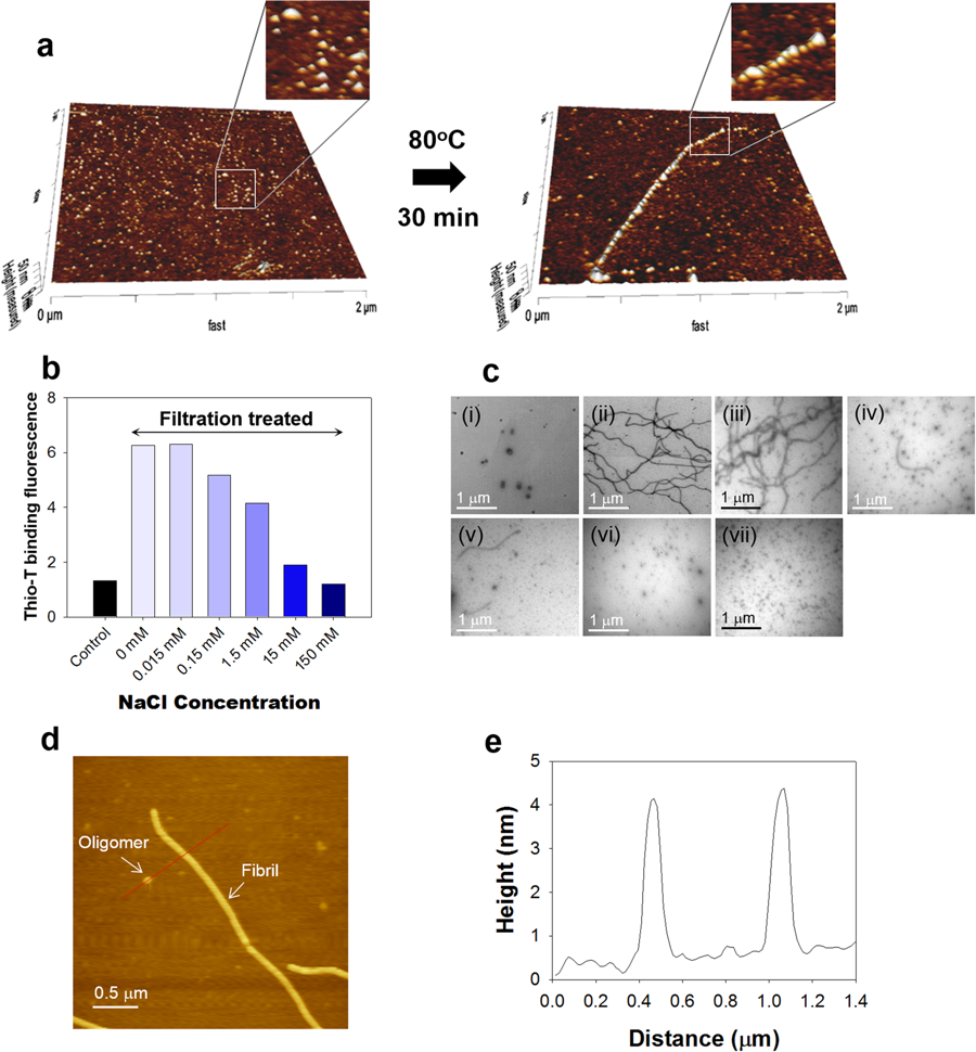
Figure 2 from Ghibom Bhak et al., “Morphological Evaluation of Meta-stable Oligomers of α-Synuclein with Small-Angle Neutron Scattering”:
Unit-assembly of Meta-αS-Os. (a) 3-D AFM images of the scattered Meta-αS-Os species and an extended fibrillar structure on the mica surface. Black arrow represents the heat treatment at 80 °C for 30 min. (b) Thio-T binding fluorescence of αS after the centrifugal membrane filtration in the absence or presence of NaCl at various concentrations of 0, 0.015, 0.15, 1.5, 15, and 150 mM. (c) TEM images of the αS oligomers before (i) and after the membrane filtration at various concentrations of 0 (ii), 0.015 (iii), 0.15 (iv), 1.5 (v), 15 (vi) or 150 mM (vii). (d) AFM image showing coexistence of the oligomers and the amyloid fibrils at 1.5 mM NaCl following the membrane filtration. (e) AFM height profile of the oligomeric and fibrillar species.
“For the assessment of heat-induced oligomeric unit assembly, an aliquot (10 μl) containing αS oligomers was placed on a cleaved mica surface. Following 30-min incubation at room temperature, the mica was immersed in fresh 20 mM Mes (pH 6.5) and incubated at 80 °C for another 30 min. After the heat-treated mica was cleaned with excessive distillated water, the oligomeric and fibrillar species of αS on the mica were analyzed with AFM in a tapping mode with a Super Sharp AFM tip (SSS-NCHR, NANOSENSORS, Switzerland). To reveal coexistence of the oligomers and fibrils, the αS species (10 μl) was adsorbed on the mica coated with poly-l-lysine for 5 min at room temperature. After the mica was washed with distilled water 5 times and dried in vacuum chamber, AFM analysis was done in a tapping mode.”*
“Meta-αS-Os have played a growing unit for the unit-assembly-based facilitated amyloid fibril formation. Their unit-assembly was examined on the surface of mica immersed in 20 mM Mes (pH 6.5) at 80 °C for 30 min. In the 3-D AFM images, the scattered Meta-αS-Os on the mica surface became aligned with each other to form the extended fibrils following the heat treatment (Fig. 2a). This phenomenon reflects general two-step process of protein-surface interaction showing initial reversible binding of the oligomers to the surface followed by their irreversible stabilization into the amyloid fibrils via the transition of Meta-αS-Os into type-Bon oligomers upon the heat treatment. […] AFM analysis revealed that the particulates and the short fibrils of αS obtained with 1.5 mM NaCl had almost identical height profile with the maximum height of 4 nm (Fig. 2d,e).”*
[…]
“AFM analysis revealed that the particulates and the short fibrils of αS obtained with 1.5 mM NaCl had almost identical height profile with the maximum height of 4 nm (Fig. 2d,e).”*
*Ghibom Bhak, Soonkoo Lee, Tae-Hwan Kim, Ji-Hye Lee, Jee Eun Yang, Keehyoung Joo, Jooyoung Lee, Kookheon Char and Seung R. Paik
Morphological Evaluation of Meta-stable Oligomers of α-Synuclein with Small-Angle Neutron Scattering
Nature Scientific Reports, volume 8, Article number: 14295 (2018)
DOI: https://doi.org/10.1038/s41598-018-32655-0
Open Access: The article “Morphological Evaluation of Meta-stable Oligomers of α-Synuclein with Small-Angle Neutron Scattering” by Ghibom Bhak et al. is licensed under a Creative Commons Attribution 4.0 International License, which permits use, sharing, adaptation, distribution and reproduction in any medium or format, as long as you give appropriate credit to the original author(s) and the source, provide a link to the Creative Commons license, and indicate if changes were made. The images or other third party material in this article are included in the article’s Creative Commons license, unless indicated otherwise in a credit line to the material. If material is not included in the article’s Creative Commons license and your intended use is not permitted by statutory regulation or exceeds the permitted use, you will need to obtain permission directly from the copyright holder. To view a copy of this license, visit http://creativecommons.org/licenses/by/4.0/.