The dielectric permittivity of membranes is important for many fundamental electrophysiological functions like selective transport in ion channels, action potential propagation and energy generation.*
In their article “Nanoscale dipole dynamics of protein membranes studied by broadband dielectric microscopy” George Gramse, Andreas Schönhals and Ferry Kienberger investigate the nearfield dipole mobility of protein membranes in a wide frequency range from 3 kHz to 10 GHz.*
They achieved their results by adding the frequency as a second fundamental dimension to quantitative dielectric microscopy thereby demonstrating the possibilities of broadband dielectric microscopy for the investigation of dynamic processes in cell bioelectricity at the individual molecular level. Furthermore, the technique may also shed light on local dynamic processes in related materials science applications like semiconductor research or nano-electronics.*
All AFM measurements were carried out at 25 °C using a NANOSENSORS Platinum Silicide AFM probe ( PtSi-FM ).
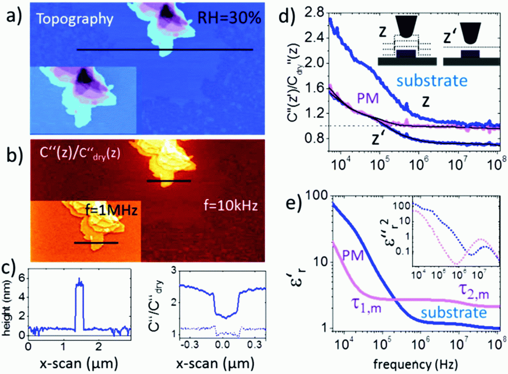
Fig. 2 from “Nanoscale dipole dynamics of protein membranes studied by broadband dielectric microscopy” by Gramse et al.: (a) AFM topography and (b) corresponding C′′(z)/C′′dry(z) image obtained in lift mode at z = 10 nm above the last scan line and at a frequency of ω = 10 kHz (inset at 1 MHz). The corresponding topography and C′′(z)/C′′dry(z) profile lines are shown in (c). Solid lines correspond to profile lines at 10 kHz and the dashed line to 1 MHz. (d) Normalized dielectric spectra on the substrate and protein membrane at constant height z′ = 15 nm and lift mode z = 15 nm. Black solid lines represent fitting with eqn (1) and (2). (e). Resulting complex dielectric functions ε′r(f) and ε′′r(f)2 (using the relation ε′′r(f) = −(π/2∂)ε′r/∂ln(2πf)38).
All measurements are carried out at 25 °C using PtSi-FM tips from NANOSENSORS (Germany). Humidity was changed and left to stabilize for 2–3 hours. Imaging conditions were adjusted to maintain the lift distance for the dielectric images identical.
*Georg Gramse, Andreas Schönhals, Ferry Kienberger
Nanoscale dipole dynamics of protein membranes studied by broadband dielectric microscopy
Nanoscale, 2019, 11, 4303-4309
DOI: 10.1039/C8NR05880F
Please follow this external link for the full article: https://pubs.rsc.org/en/content/articlehtml/2019/nr/c8nr05880f
Open Access The article “Nanoscale dipole dynamics of protein membranes studied by broadband dielectric microscopy” by George Gramse, Andreas Schönhals and Ferry Kienberger is licensed under a Creative Commons Attribution 3.0 International License, which permits use, sharing, adaptation, distribution and reproduction in any medium or format, as long as you give appropriate credit to the original author(s) and the source, provide a link to the Creative Commons license, and indicate if changes were made. To view a copy of this license, visit https://creativecommons.org/licenses/by/3.0/