Xia Liu, Amit Kumar Sachan, Samuel Tobias Howell, Ana Conde-Rubio, Armin W. Knoll, Giovanni Boero, Renato Zenobi, and Jürgen Brugger
Thermomechanical Nanostraining of Two-Dimensional Materials
Nano Lett. 2020, 20, 11, 8250–8257
DOI: https://doi.org/10.1021/acs.nanolett.0c03358
Xiaolong Kou, Qian Zhou, Dong Wang, Jinghe Yuan, Xiaohong Fang, Lijun Wan
High-resolution imaging of graphene by tip-enhanced coherent anti-Stokes Raman scattering
Journal of Innovative Optical Health SciencesVol. 12, No. 01, 1841003 (2019)
https://doi.org/10.1142/S1793545818410031
https://www.worldscientific.com/doi/full/10.1142/S1793545818410031
Andrea Mancini, Valeria Giliberti, Alessandro Alabastri, Eugenio Calandrini, Francesco De Angelis , Denis Garoli, and Michele Ortolani
Thermoplasmonic Effect of Surface-Enhanced Infrared Absorption in Vertical Nanoantenna Arrays
J. Phys. Chem. C 122 (2018), 24, 13072-13081
DOI: 10.1021/acs.jpcc.8b03808
https://pubs.acs.org/doi/abs/10.1021/acs.jpcc.8b03808
https://arxiv.org/ftp/arxiv/papers/1801/1801.07465.pdf
A. Bhattaraia, P. Z. El-Khoury
Imaging localized electric fields with nanometer precision through tip-enhanced Raman scattering
Chem. Commun., 2017,53, 7310-7313
DOI: 10.1039/C7CC02593A
http://pubs.rsc.org/en/content/articlelanding/2017/cc/c7cc02593a/unauth#!divAbstract
Emilie Sakat, Valeria Giliberti, Monica Bollani, Andrea Notargiacomo, Marialilia Pea, Marco Finazzi, Giovanni Pellegrini, Jean-Paul Hugonin, Alexander Weber-Bargioni, Mauro Melli, Simone Sassolini, Stefano Cabrini, Paolo Biagioni, Michele Ortolani, and Leonetta Baldassarre
Near-Field Imaging of Free Carriers in ZnO Nanowires with a Scanning Probe Tip Made of Heavily Doped Germanium
Phys. Rev. Applied 8, 054042 – Published 21 November 2017
DOI: https://doi.org/10.1103/PhysRevApplied.8.054042
https://hal-iogs.archives-ouvertes.fr/hal-01698535/document
Ashish Bhattarai, Alan G. Joly, Wayne P. Hess , Patrick Z. El-Khoury
Visualizing Electric Fields at Au(111) Step Edges via Tip-Enhanced Raman Scattering
Nano Lett., 2017, 17 (11), pp 7131–7137
DOI: 10.1021/acs.nanolett.7b04027
https://pubs.acs.org/doi/abs/10.1021/acs.nanolett.7b04027
Ewelina Lipiec, Aleksandre Japaridze, Jacek Szczerbiński, Giovanni Dietler, Renato Zenobi
Preparation of Well‐Defined DNA Samples for Reproducible Nanospectroscopic Measurements
Small, Volume12, Issue35, September 21, 2016, Pages 4821-4829
https://doi.org/10.1002/smll.201601711
https://onlinelibrary.wiley.com/doi/abs/10.1002/smll.201601711
Aleksandre Japaridze, Dusan Vobornik, Ewelina Lipiec, Andrea Cerreta, Jacek Szczerbinski, Renato Zenobi, and Giovanni Dietler
Toward an Effective Control of DNA’s Submolecular Conformation on a Surface
Macromolecules, 2016, 49 (2), pp 643–652
DOI: 10.1021/acs.macromol.5b01827
https://pubs.acs.org/doi/abs/10.1021/acs.macromol.5b01827
https://www.researchgate.net/profile/Dusan_Vobornik/publication/290474779_Toward_an_Effective_Control_of_DNA%27s_Submolecular_Conformation_on_a_Surface/links/56a7807c08ae0fd8b3fe0a03/Toward-an-Effective-Control-of-DNAs-Submolecular-Conformation-on-a-Surface.pdf
Lu, P., Li, J., Wang, D. et al. Sci. China Chem. (2015) 58: 1494.
Si@Cu@Au AFM tips for tip-enhanced Raman spectrum
doi: 10.1007/s11426-015-5353-6,
http://link.springer.com/article/10.1007/s11426-015-5353-6
Mónica Sánchez-Román, David Fernández-Remolar, Ricardo Amils, Antonio Sánchez-Navas, Thomas Schmid, Patxi San Martin-Uriz, Nuria Rodríguez, Judith A. McKenzie, Crisogono Vasconcelos (2014), Microbial mediated formation of Fe-carbonate minerals under extreme acidic conditions
Scientific Reports, Volume 4, 4767,
DOI: 10.1038/srep04767
http://dx.doi.org/10.1038/srep04767
http://www.nature.com/articles/srep04767
Christoph Huber ac, Andreas Trügler b, Ulrich Hohenester b, Yehiam Prior c and Wolfgang Kautek (2013)
Optical near-field excitation at commercial scanning probe microscopy tips: a theoretical and experimental investigation
Phys. Chem. Chem. Phys., 2014, 16, 2289-2296
DOI: 10.1039/C3CP51730F
http://pubs.rsc.org/en/content/articlehtml/2013/cp/c3cp51730f
Ian M. Craig ; Mark C. Phillips ; Matthew S. Taubman ; Erik E. Josberger ; Markus B. Raschke (2013),
Infrared scattering scanning near-field optical microscopy using an external cavity quantum cascade laser for nanoscale chemical imaging and spectroscopy of explosive residues
Proc. SPIE 8631, Quantum Sensing and Nanophotonic Devices X, 863110 (February 4, 2013)
doi: 10.1117/12.2004954
http://proceedings.spiedigitallibrary.org/proceeding.aspx?articleid=1568446
Melissa Paulite, Carolin Blum, Thomas Schmid, Lothar Opilik, Klaus Eyer, Gilbert C. Walker, and Renato Zenobi
Full Spectroscopic Tip-Enhanced Raman Imaging of Single Nanotapes Formed from β-Amyloid(1–40) Peptide Fragments
ACS Nano 7, 2, 911-920 (2013)
DOI: 10.1021/nn305677k
https://pubs.acs.org/doi/abs/10.1021/nn305677k
Asghari-Khiavi, M., Wood, B. R., Hojati-Talemi, P., Downes, A., McNaughton, D. and Mechler, A. (2012),
Exploring the origin of tip-enhanced Raman scattering; preparation of efficient TERS probes with high yield.
J. Raman Spectrosc., 43: 173–180.
doi: 10.1002/jrs.3021
http://onlinelibrary.wiley.com/doi/10.1002/jrs.3021/full
Blum, C., Schmid, T., Opilik, L., Weidmann, S., Fagerer, S. R. and Zenobi, R. (2012),
Understanding tip-enhanced Raman spectra of biological molecules: a combined Raman, SERS and TERS study.
J. Raman Spectrosc., 43: 1895–1904.
doi: 10.1002/jrs.4099
http://onlinelibrary.wiley.com/doi/10.1002/jrs.4099/full
R. Ramos, M. J. Gordon (2012),
Near-field artifacts in tip-enhanced Raman spectroscopy,
Applied Physics Letters , Volume 100, Issue 21 ,
DOI: http://dx.doi.org/10.1063/1.4722805
http://aip.scitation.org/doi/abs/10.1063/1.4722805
K. L. Andrew Chan, Sergei G Kazarian (2011),
Tip-enhanced Raman mapping with top-illumination AFM,
Nanotechnology, Volume 22, Number 17 (mentioning NTEGRA Spectra and ATEC)
http://iopscience.iop.org/article/10.1088/0957-4484/22/17/175701/meta
Roman M. Balabin, Rustem Z. Syunyaev, Thomas Schmid, Johannes Stadler, Ekaterina I. Lomakina, and Renato Zenobi (2011),
Asphaltene Adsorption onto an Iron Surface: Combined Near-Infrared
(NIR), Raman, and AFM Study of the Kinetics, Thermodynamics, and Layer Structure
Energy & Fuels 2011 25 (1), 189-196,
DOI: 10.1021/ef100779a (mentioning NTEGRA Spectra and ATEC)
https://www.researchgate.net/publication/228394305_Asphaltene_Adsorption_onto_an_Iron_Surface_Combined_Near-Infrared_NIR_Raman_and_AFM_Study_of_the_Kinetics_Thermodynamics_and_Layer_Structure
Przemyslaw R. Brejna, Peter R. Griffiths (2010),
Electroless Deposition of Silver onto Silicon as a Method of Preparation of Reproducible Surface-Enhanced Raman Spectroscopy Substrates and Tip-Enhanced Raman Spectroscopy Tips, http://journals.sagepub.com/doi/pdf/10.1366/000370210791211682
Peica, N., Röhrig, S., Rüdiger, A., Brose, K., Thomsen, C. and Maultzsch, J. (2009),
Characterization of dye molecules and carbon nanostructures by tip-enhanced Raman spectroscopy.
Phys. Status Solidi B, 246: 2708–2712.
doi:10.1002/pssb.200982278
http://onlinelibrary.wiley.com/doi/10.1002/pssb.200982278/full
Poborchii, V., Tada, T., Kanayama, T. and Geshev, P. (2009),
Optimization of tip material and shape for near-UV TERS in Si structures.
J. Raman Spectrosc., 40: 1377–1385.
doi: 10.1002/jrs.2417
https://www.researchgate.net/profile/Pavel_Geshev/publication/230006436_Optimization_of_tip_material_and_shape_for_near-UV_TERS_in_Si_structures/links/02e7e51dd963447729000000.pdf
A. Ono, K Masui, Y Saito, T Sakata, A Taguchi et. Al. (2008),
Active Control of the Oxidization of a Silicon Cantilever for the Characterization of Silicon-based Semiconductors,
Chemistry Letters, Vol. 37, No.1, 122-123,
DOI: http://dx.doi.org/10.1246/cl.2008.122
http://citeseerx.ist.psu.edu/viewdoc/download?doi=10.1.1.669.9556&rep=rep1&type=pdf
Y. Saito, M. Motohashi, and N. Hayazawa M. Iyoki S. Kawata (2006),
Nanoscale characterization of strained silicon by tip-enhanced Raman spectroscope in reflection mode,
Applied Physics Letters, Volume 88, Issue 14,
DOI: http://dx.doi.org/10.1063/1.2191949
http://aip.scitation.org/doi/abs/10.1063/1.2191949


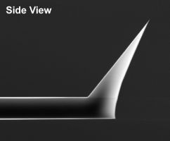
 Due to their unique geometry the tips of the AdvancedTEC probes are more susceptible to tip damage by electrostatic discharge (ESD) than other Silicon-SPM-Probes.


